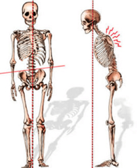
Back Bones Diagram / Exercises can strengthen the core muscles that support the spine and.. Vertebrae are the structural constituents of the spine.there are 33 vertebrae in total; The occiput (co), also known as the occipital bone, is a flat bone that forms the back of the head. Lateral labeled diagram of the human vertebral spinal column showing vertebrae numbering order and the 5 different regions of the spine. The first seven bones (vertebrae) of your spine form your neck. Seven cervical vertebrae in the neck, twelve thoracic vertebrae in the torso and five lumbar vertebrae in the lower back.
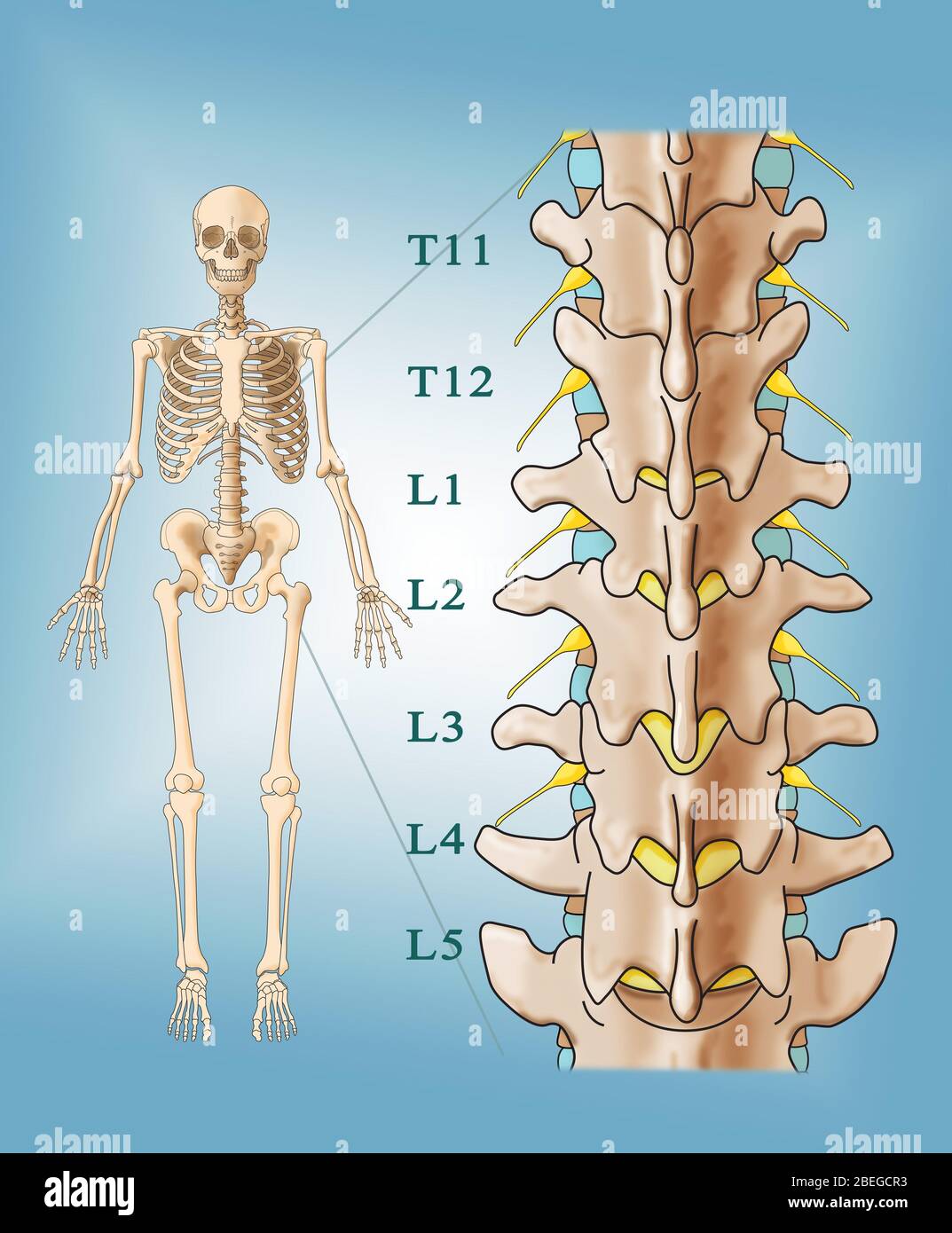
Bone diagram forehead (frontal bone) nose bones (nasals) cheek bone (zygoma) upper jaw (maxilla) lower jaw (mandible) breast bone (sternum). Each lumbar spinal level is numbered from top to bottom—l1 through l5, or l6. The first seven bones (vertebrae) of your spine form your neck. Key parts of your spine include vertebrae (bones), disks, nerves and the spinal cord. The disks that cushion vertebrae may compress with age or injury, leading to a herniated disk.
Human Body Anatomy Skeleton With Veins And Stock Illustration 57700553 Pixta from en.pimg.jp Its appearance is different from the other spinal vertebrae. 12 photos of the human back bone chart. The l5 vertebra is connected to the top of. Daniel nelson on january 1, 2019 2 comments 🔥! (temporal bone) shoulder blade (scapula) lower back vertebrae (5) (lumbar vertebrae) back of skull (occipital bone) fused vertebrae (5) (sacrum) hand bones (metacarpals) finger bones It consists of 5 lumbar vertebra that are numbered 1 through 5 from top to bottom i.e. The first seven bones (vertebrae) of your spine form your neck. The spine or backbone consists of 26 small bones or vertebrae.
They help support particular bones and make them move.
This human anatomy module is composed of diagrams, illustrations and 3d views of the back, cervical, thoracic and lumbar spinal areas as well as the various vertebrae. The disks that cushion vertebrae may compress with age or injury, leading to a herniated disk. L1, l2, l3, l4, and l5. This spinal column provides the main support for your body, allowing you to stand upright, bend, and twist, while protecting the spinal cord from injury. It is also known as the vertebral column. Spine diagram studying a spine diagram is one way to better understand many of the individual components of the back bone and how they might relate to a symptomatic back, neck or sciatica pain condition. Spinal vertebrae bone spine vertebra toracica spinal cord spine structure back diagram spine sections spinal cord vertebrae spinal structure health diagram. The lumbar spine makes up the the lower end of the spinal column. It connects with the collarbone at the front of the body. Exercises can strengthen the core muscles that support the spine and. Each typical vertebra consists of a body, an arch and three processes that stem from. 12 photos of the human back bone chart. Bone diagram forehead (frontal bone) nose bones (nasals) cheek bone (zygoma) upper jaw (maxilla) lower jaw (mandible) breast bone (sternum).
The first seven bones (vertebrae) of your spine form your neck. 12 photos of the human back bone chart. These bones are connected at the back with specialized joints. The disks that cushion vertebrae may compress with age or injury, leading to a herniated disk. Daniel nelson on january 1, 2019 2 comments 🔥!
Back And Spine Complete Pain Care Helping You Return To You from www.completepaincare.com The spine anatomy is a complex structure. L1, l2, l3, l4, and l5. The muscles, bones, ligaments, and tendons in the back can all be injured and cause back. Exercises can strengthen the core muscles that support the spine and. This human anatomy module is composed of diagrams, illustrations and 3d views of the back, cervical, thoracic and lumbar spinal areas as well as the various vertebrae. The spine is made of 33 individual bones stacked one on top of the other. Bones of the pelvis and lower back. The atlas is a ring of bone made up of two lateral masses joined at.
The bones of the pelvis and lower back work together to support the body's weight, anchor the abdominal and hip muscles, and protect the delicate vital organs of the vertebral and abdominopelvic cavities.
The spine diagram the spine diagram shown below, consists of many bones or vertebrae,soft discs,the spinal cord, and spinal nerves. This spinal column provides the main support for your body, allowing you to stand upright, bend, and twist, while protecting the spinal cord from injury. This human anatomy module is composed of diagrams, illustrations and 3d views of the back, cervical, thoracic and lumbar spinal areas as well as the various vertebrae. This diagram depicts back skeletal anatomy with parts and labels. L1, l2, l3, l4, and l5. More commonly known as the shoulder blade, the scapula is a flat triangular bone located in the upper back. Posted in bones , diagrams | tagged body skeleton , human skeletal anatomy , human skeleton , human skeleton anatomy , skeletal , skeletal anatomy , skeletal images. Its appearance is different from the other spinal vertebrae. A tough, springy disc of cartilage sits between the vertebrae of your spine. The vertebral column is a part of the axial skeleton, which comprises the skull, ribs and sternum other than the vertebral column. Strong muscles and bones, flexible tendons and ligaments, and sensitive nerves contribute to a healthy spine. The vertebrae, which stack like spools of thread, support the back and protect the spinal cord. Your lower back contains 5 vertebral bones stacked above each other with intervertebral discs in between.
The disks that cushion vertebrae may compress with age or injury, leading to a herniated disk. The spine diagram the spine diagram shown below, consists of many bones or vertebrae,soft discs,the spinal cord, and spinal nerves. The muscles, bones, ligaments, and tendons in the back can all be injured and cause back. Strong muscles and bones, flexible tendons and ligaments, and sensitive nerves contribute to a healthy spine. This spinal column provides the main support for your body, allowing you to stand upright, bend, and twist, while protecting the spinal cord from injury.
Anatomy Of Lumbar Spine High Resolution Stock Photography And Images Alamy from c8.alamy.com Vertebrae are the structural constituents of the spine.there are 33 vertebrae in total; The bones of the pelvis and lower back work together to support the body's weight, anchor the abdominal and hip muscles, and protect the delicate vital organs of the vertebral and abdominopelvic cavities. This diagram depicts back skeletal anatomy with parts and labels. It is particularly interesting for physiotherapists. Related posts of human back bones diagram bone structure birds. Its appearance is different from the other spinal vertebrae. Powerful muscles that move the head and arms attach to these bones as well. Muscle or tendon injuries can occur anywhere in the body.
Bone structure birds 12 photos of the bone structure birds bone structure birds, bone structure in.
It also covers some common conditions and injuries that can affect the back. Its appearance is different from the other spinal vertebrae. The spine anatomy is a complex structure. The occiput (co), also known as the occipital bone, is a flat bone that forms the back of the head. The bones of the chest and upper back combine to form the strong, protective rib cage around the vital thoracic organs such as the heart and lungs. Bones of the pelvis and lower back. The spine is made of 33 individual bones stacked one on top of the other. The atlas is a ring of bone made up of two lateral masses joined at. A tough, springy disc of cartilage sits between the vertebrae of your spine. This human anatomy module is composed of diagrams, illustrations and 3d views of the back, cervical, thoracic and lumbar spinal areas as well as the various vertebrae. But, they are common in the back and can cause pain. Daniel nelson on january 1, 2019 2 comments 🔥! The disks that cushion vertebrae may compress with age or injury, leading to a herniated disk.